Translating
Tacit Medical Knowledge Into Explicit Knowledge
Sachiko Iwai, Fukuya Ishino, Waseda University , Japan
ABSTRACT:
Cytological diagnosis is a method to detect cancer in which qualified specialists distinguish between cancerous and non-cancerous cells using an optical microscope. However, the medical knowledge used for diagnosis is tacit, which means that the result can vary depending on the education and experience of each qualified specialist (Tezuka F. 1991). In this study, the top 10% of specialists were first identified. Then their superior tacit knowledge was translated into explicit knowledge by the long-term collaboration of a top specialist and two amateurs who have no medical knowledge. This explicit knowledge was helpful for cytological diagnosis, even allowing amateurs to diagnose the cells. This translation of tacit knowledge into explicit knowledge allowed a manual to be created, which suggests that it may be possible to develop a computer program for cytological diagnosis.
Keywords: Cytological diagnosis, Tacit knowledge, Explicit knowledge, Diagnostic
algorithm
1. Background
Urine cytology is widely used for bladder cancer diagnosis as it is an inexpensive and pain-free technique. However, it is known that there are judgment variations on the same material among specialists depending on education and experience (Dawson et al, 1991). As the actual judgment variation and error rate have not been investigated, it is not known which specialist has acquired more correct knowledge through experience. Around the world, doctors who refer to specialists may know which are the most reliable. If the tacit knowledge which reliable specialists have acquired can be transmitted to other less experienced specialists, it should lead to decreases in both unnecessary operations and delays in the detection of cancer.
2. Methods
In order to translate excellent tacit knowledge into explicit knowledge which can be propagated to other specialists, the following five steps were undertaken.
1. Survey the variation in the specialists’ judgments
2. Identify the top-ranking specialists
3. Extract the decision knowledge of the top-ranking specialists
4. Write the manual
5. Verify the manual by evaluating diagnosis by amateurs
These five steps will now be described in detail.
Step I: Survey The Variation In The
Specialists’ Judgments
Before the survey, photographs of 120 cells and the correct diagnoses were prepared from past cases. The 120 photographs were delivered to 46 specialists working in 15 hospitals nationwide who had agreed to participate in the study. The correct answers were not sent. The participants were also asked to rank 10 common cell characteristics in terms of their usefulness for cytological diagnosis. The responses returned contained the diagnosis of ‘benign’, ‘malignant’ or ‘borderline’ for each of the 120 cells, and the priority levels assigned to the 10 characteristics. Because the survey could be viewed as a test of judgment abilities, anonymity was promised in order to prevent any loss of reputation by the participants. Thus, the test data consists of the survey data of 120×46 responses and a set of 120 correct answers.
Step II: Identify The Top-Ranking Specialists
The answers from the 46 participants can be arranged as the matrix

In the matrix, the entry Dij represents the answer given for i-th cell by j-th cooperator: it is set to +1 for ‘malignant’, 0 for ‘borderline’ and -1 for ‘benign’. The correct answer for the i-th cell, Ti , is also assigned a value of +1, 0, or -1 using the same scale. The true state of the cell (correct answers) can then be arranged as the matrix
. 
Then, the judgment error in each case, Eij is given by
Eij = Dij -
Ti. (1)
Therefore, the error matrix is

where Eij
takes a value out of {+2, +1, 0, -1, -2}. The values of Eij can be interpreted as follows:
+2: A benign cell is diagnosed as malignant.
+1: A benign cell is diagnosed as borderline, or a borderline cell is diagnosed
as malignant,
0: Correct diagnosis.
-1: A malignant cell is diagnosed as borderline, or a borderline cell is
diagnosed as benign,
-2: A malignant cell is diagnosed as benign. This is a risky judgment which
will delay cancer detection.
Reliable specialists are those who fulfill the following 4 conditions:
a) Their concordance ratio (correct answer rate) is high.
b)
Their root mean square (RMS) error, REj, given by

is small.
c) Their answers do not include the most serious errors (Eij = -2).
d) They do not give ‘borderline’ answers unnecessarily.
The RMS errors of the 46 specialists are shown in
Figure 1.These errors are highly correlated with the specialists’
concordance ratios (see Figure 2).
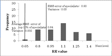
Figure 1: Bar Chart Of Root Mean Square Error
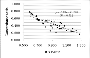
Figure 2: Relationship Between Concordance Ratio And
RMS Error
Δ Indicates Specialists In
The Top 10%.
The top 10% of specialists (5 participants) were determined by concordance ratio ranking. From Figure 2 it can be seen that if the concordance ratio of a specialist is good (high), their RMS error is also good (low). Then, focusing on this top 10% of specialists, the other 2 conditions were reviewed. From Table 1 it can be seen that the judgments of these specialists do not include ‘dangerous’ errors (Eij = -2) and ‘borderline’ decisions are fewer than average. As the top 10% of specialists have bee identified, the next step is to codify their knowledge.
Table 1: Review Of 4 Conditions For Top 10% Of
Specialists
|
Concordance ratio rank |
Specialists
respondent number |
Concordance
ratio |
RE |
Rank by RE |
Number of Eij
= −2 |
Number of Eij
= +2 |
Number
of Dij = 0 |
|
1 |
9 |
0.78 |
0.59 |
1 |
0 |
5 |
14 |
|
2 |
14 |
0.73 |
0.61 |
2 |
0 |
4 |
24 |
|
3 |
32 |
0.71 |
0.63 |
3 |
0 |
4 |
22 |
|
4 |
25 |
0.67 |
0.68 |
4 |
0 |
4 |
20 |
|
5 |
16 |
0.59 |
0.70 |
6 |
0 |
3 |
37 |
|
Average
of specialists |
|
0.44 |
0.90 |
|
10 |
1 |
57 |
Step III: Extract The Decision Knowledge Of The
Top-Ranking Specialists
First, the priority of rules used for diagnosis rules by the top-ranking specialists must be distinguished from those used by low ranked specialists. Once this is done, the knowledge must be translated into explicit knowledge which is understandable even for beginners or amateurs (Step IV).
The following 10 items are
descriptions of the characteristics that specialists were found to use in
cytological diagnosis. The descriptions of the characteristics are not only too
difficult for lay people to understand but also include ambiguous expressions
whose meaning may sometimes vary depending on experience. The medical jargon
and ambiguous words are given in italics.
a)
Cell binding * In general, benign
cells in urine appear as single cells or as several
cells, and are rarely observed in groups. After calculus
or endoscopic treatment, large groups of strongly
binding cells may appear. In malignant cells, there is a
tendency for single cells to be weakly binding and appear in
groups. Sometimes, groups consisting of over 30 cells may appear
which do not appear with benign cells.
b)
Cell size * Although
care is required in the case of extremely large cells or
significant disparities in cell size, enlargement is
commonly observed due to inflammation*, calculus*,
and cancer therapy (chemotherapy* and radiotherapy*). Although malignancy may
be considered if the size is more than 6 times larger than white blood
cells*, large cells are not limited to malignancy, and may be
benign even if the entire cell is large as long as the nucleus is small. Small
cells* where the nucleus is large may be cancer or dysplasia.
c)
N/C ratio* The N/C ratio is the
area ratio of the nucleus to cytoplasm in the cell, and increases in
malignant cells. If the area ratio of nucleus to cytoplasm is greater
than 0.5, the possibility of malignancy is considered. Although the
possibility of malignancy increases as the N/C ratio increases, the possibility
of malignancy is extremely high for area ratios of over 0.8. In bare nucleus
cells* that only exhibit a nucleus and do not have a cytoplasm, this is not
conclusive. The area of the nucleus may be large because of degeneration*
or reactivity* with viral infections* or treatment.
d)
Location of the nucleus* Although the position
of the nucleus in the cells appearing in urine is often eccentric,
in malignant cells the eccentricity is often extreme.
e)
Atypical nuclear shape* Although the nucleus in
benign cells is circular or elliptical, in
cases where there is a large amount of undulations or solid
cutouts, the possibility of malignancy is high.
f)
Amount of nuclear chromatin* This indicates the density
of chromatin. The nuclear structure is confirmed, and
cells that exhibit deep dye-affinity for dark purple colors compared
to the background white blood cells have a possibility of malignancy. Regardless
of whether malignant or benign, care is required because the terminal
state of cellular degeneracy is concentration
of the nucleus, which exhibits deep dye affinity.
g)
Nuclear chromatin distribution* In new cells and
cells that have degenerated, the chromatin state has changed. Cells
that have a variety of chromatin granule sizes corresponding to a
mixture of thick and thin chromatin networks* have a high
possibility of being malignant cells that have degenerated. Although new cells
that feel non-transparent and exhibit large amount of euchromatin*
that appears like salt and pepper do not exhibit deep dye-affinity in the
nucleus and have a high possibility of malignancy, new cells are hard to obtain
in normal testing and euchromatin is not commonly found.
h)
Nuclear envelope irregular thickening* Indicates a state where
chromatin is adhered to the nuclear membrane*. In vivid
cancer cells, the nuclear envelope* is thin as if drawn
by a sharp pencil, and is difficult to visually distinguish* from
benign cells. In degenerate cancer cells, the thickness of the
nuclear envelope is irregular, as if drawn by a pencil where the tip has
been cut into a square*, and areas with significant thickness can also
be observed.
i)
Nuclear protrusions* In malignant cells, DNA
activity* is heightened, and the nucleus becomes solid
and can be observed to protrude from the cytoplasm.
j)
Nucleolus* In malignant cells,
although nucleolus growth* or increases in number*
may be observed because RNA activity* is also heightened, this
often cannot be confirmed in malignant cells with strong degeneration.
Specialists were asked to rate these ten cell characteristics. Three points were given for characteristics that are always important and two points for those that can be important depending on the case. One point was given for characteristics that are not important even if found. The importance of the various characteristics were then categorized according to the total number of evaluation points Tables 2-4 show priorities of the top 10% of specialists, those of the bottom 10% of specialists and the overall priorities.
Table 2: Ranking Of The Importance Of The 10 Cell
Characteristics By The Top 10% Of Specialists
|
Rank |
Cell
Characteristic |
||
|
1 |
N/C
ratio |
Chromatin
quantity |
Nuclear
protrusions |
|
2 |
Atypical
nuclear shape |
Cell
size |
|
|
3 |
Cell
binding |
Location
of nucleus |
|
|
4 |
Chromatin
distribution |
Nuclear
envelope irregular thickening |
|
|
5 |
Nucleolus |
|
|
Table 3: Ranking Of The Importance
Of The 10 Cell Characteristics By The Bottom 10% Of Specialists
|
Rank |
Cell
Characteristic |
||
|
1 |
Chromatin
quantity |
Nuclear
protrusions |
|
|
2 |
N/C
ratio |
Cell
size |
|
|
3 |
Cell
binding |
Chromatin
distribution |
Nuclear envelope irregular thickening |
|
4 |
Location
of nucleus |
Atypical
nuclear shape |
|
|
5 |
Nucleolus |
|
|
Table 4: Specialists Overall Importance Rankings Of
The 10 Cell Characteristics
|
Rank |
Cell
Characteristic |
||
|
1 |
N/C
ratio |
Chromatin
quantity |
|
|
2 |
Nuclear
protrusions |
Atypical
nuclear shape |
|
|
3 |
Cell
binding |
Location
of nucleus |
Cell
size |
|
4 |
Chromatin
distribution |
Nuclear
envelope irregular thickening |
|
|
5 |
Nucleolus |
|
|
The final priority order given in Table 5 was determined by the following rules:
1.
Take the responses of the top 10% of specialists
and order the characteristics by the total number of evaluation points (see Table
2). The ties in Table 2 were resolved by applying rules 2 and 3.
2.
For characteristics given the same ranking by
the top 10% of specialists, those that were less emphasized by the bottom 10%
of specialists (see Table 3) were given the highest rankings.
If two characteristics were also given equal rank by the
bottom 10% of specialists, the overall importance score assigned by all the
specialists was taken into account. The characteristic with the higher overall
score was ranked higher in the final table.
For example, the N/C ratio placed first in the final table
because the N/C ratio was viewed as important by the top-ranking specialists
and evaluated as less important by the bottom ranking specialists. Because
there is no difference between chromatin quantity and nuclear protrusions in
Table 3, the overall rankings (Table 4) were considered and the chromatin
quantity, which had more points, was selected as the second rank
characteristic. The third rank characteristic was thus determined to be the presence
of nuclear protrusions. We now return to Table 3 and focus on the second ranked
characteristics of atypical nuclear shape and cell size. By proceeding in this
way, the tacit knowledge of the top 10% of specialists was elicited to decide
on the priorities given in Table 5.
Table 5: The Final Priorities For Diagnosis Rules.
Cell With The Same Color Density
Are Characteristics With The Same Importance Level For The Top 10% Of
Specialists As Shown In Table 2.
|
Priority |
Cell
Characteristics |
|
1 |
N/C Ratio |
|
2 |
Chromatin
Quantity |
|
3 |
Nuclear
Protrusions |
|
4 |
Atypical
Nuclear Shape |
|
5 |
Cell
Size |
|
6 |
Location
of Nucleus |
|
7 |
Cell
Binding |
|
8 |
Chromatin
Distribution |
|
9 |
Nuclear
Envelope Irregular Thickening |
|
10 |
Nucleolus |
Step IV: Write The Manual
Translating the 10 rules into
explicit knowledge needs patience because they include not only medical jargon
but also ambiguous words, such as ‘big’, ‘many’,
‘dark’, ’round’ and
‘deformed’. The work
was done by collaboration between a top-ranking specialist and two graduate
students specializing in computer science. The specialist was responsible for
ensuring that the translated sentences did not include incorrect descriptions.
The students ensured that the descriptions were understandable, even to
amateurs. The translated sentences went back and forth until the two amateurs
could use the rules for cell diagnosis. Ambiguous words were defined by digital
expression and sometimes sample photos were used to define colors.
A diagnostic flowchart (see
Figure 3) was created for amateur participants to do the same work as the 46
specialists had done. The flowchart is a manual for diagnosing bladder cells
from microscope photos. Of course, the exits of the flowchart give benign cells, malignant cells or
borderline cells. The 10 rules appear in the order given in Table 5 (note that
rule 1 has been split into two parts).
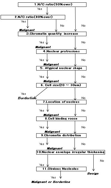
Figure 3: Flow Chart For Cytological Diagnosis
Step V: Verify The Manual By Evaluating Diagnosis
By Amateurs
In order to investigate the degree to which the manual transmits the tacit knowledge of the top-ranking specialists, the following experiment was performed. Fifty amateur test subjects (31 females and 19 males) were selected. The test subjects had the following careers, and were all thought to be capable of rational thinking.
¨ University students majoring in humanities: 3 people (2 female, 1 male)
¨ University students majoring in science: 3 people (1 female, 2 male)
¨ Graduate students studying engineering: 3 people (0 female, 3 male)
¨ Company employees in their 20s: 6 people (4 female, 2 male)
¨ Company employees in their 30s: 7 people (5 female, 2 male)
¨ Company employees in their 40s, teaching professionals: 5 people (4 female, 1 male)
¨ Company employees in their 50s, teaching professionals: 22 people (15 female, 7 male)
¨ Company employees in their 70s: 1 person (male)
The concordance ratio and RE were calculated in the same way as for the specialists. The judgment results are shown in Table 6 and Figure 4.
Table 6: Judgment Results Of Specialists Vs Amateurs
|
|
RE |
Concordance ratio |
Number judged as borderline |
|
Specialists’ average |
0.90 |
0.44 |
57 |
|
Top 10% of specialists |
0.64 |
0.69 |
26 |
|
Amateurs’ average |
0.74 |
0.72 |
13 |
|
Top 10% of amateurs |
0.60 |
0.80 |
10 |
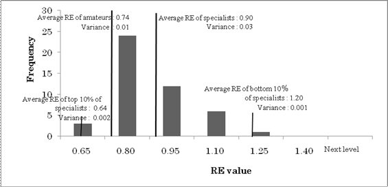
Figure 4: Bar Chart Of The Amateurs’ RMS Error
The RE distributions of both
the specialists and the amateurs were not significantly different from a normal
distribution (χ2 goodness of fit test). Both the mean and
variance of judgments (by tacit knowledge) of the top-ranking specialists were
superior. However, although the average of the amateurs’ judgments using
explicit knowledge does not surpass that of the original top-ranking
specialists, the judgment is clearly superior to that of the remaining 90% of
specialists (n=41) who depended only on tacit knowledge. Furthermore, high
negative correlations of -0.828 and -0.841 were observed between the
concordance ratio and RE for the specialists and amateurs, respectively. In
other words, it can be said that there were few participants with high
concordance ratios who made highly dangerous (Eij = -2) judgments among both the specialists and the
amateurs.
Welch’s t-test was used to perform a comparison between the specialists and amateurs based on 7 cases of dysplasia where lesions were formed (borderline judgments) and the concordance ratio of the specialists was found to be significantly higher (p < 0.05). The number of borderline judgments made by the specialists was compared to the number made by amateurs and a significant difference was found (give test & p-value). The specialists were significantly more likely to record a borderline case. Judgments of malignancy may lead to aggressive treatment and so they are important decisions. There is, therefore, a tendency toward the vague judgment of borderline among specialists. This may be the origin of this difference between the amateurs and the specialists.
3. Discussion And Conclusion
Advocates
believe that malignant cell identification is performed using comprehensive
intuition using learned knowledge and experience. By replacing the tacit
knowledge of reliable specialists with words, information that had been vague
even among specialists is arranged and a procedure was constructed for cell
diagnosis. The results obtained by amateurs based on this procedure were
extremely good. Furthermore, despite having many years’ experience in
cytological diagnosis, specialists (other than the top 10%) were worse than the
amateurs in both concordance ratio and root mean square error. This is thought
to be because of the mixture of information obtained from a large amount of
experience, with only the top-ranking specialists being able to organize this
information.
The manual created in this research was made
according to an algorithm obtained from the procedure of top-ranking
specialists for identifying malignant cells. In retrospect, this manual was
created by focusing on identifying malignant cells, and a further separate
algorithm should be investigated for cases of special lesions such as
dysplasia, which is an existing cancer state.
Cytological diagnosis is an effective method for detecting cancer and demands highly accurate diagnosis along with reproducibility. If we take reliable specialists’ views of cells to be tacit knowledge, then it is not easy to transmit this information using words to amateurs with absolutely no experience. In this paper, tacit knowledge was re-written several times into an easy yet objective representation under the supervision of amateurs familiar with computer theory. The manual that was created based on this re-writing is the result of translating tacit knowledge into explicit knowledge. This approach may lead to the development of a computer programs for diagnosis (Bacus, 1987; Jagoe & Sowter, 1984; Wolberg & Street, 1995). In recent years there has been remarkable progress in computer image recognition technology. By implementing the manual into a program, it is anticipated that high accuracy could be obtained by digitizing images and providing feedback for actual diagnosis.
4. References
Bacus, J.W. (1987), Optical Microscope System for Standardized Cell Measurements and Analysis, Applied Optimas, 26, 3280-3293.
Dawson.A.E.,Austi.,R.E.,Weinberg.D.S.(1991), Nuclear grading of breast carcinoma by image analysis: Classification by multi-variate and neural network analysis, American Journal of Clinical Pathology 95,S29-S37.
Jagoe, R., Sowter, C. (1984), Shape and Texture Analysis of Liver Cell Nuclei in Hepatomas by Computer Aided Microscopy, Journal of Clinical Pathology, 37, 755-762.
Tezuka, F. (1991), Analysis of Interobserver Reproducibility and its Improvement in Endometrial Cytology, Journal of Japanese Society of Clinical Cytology, 30(6), 1050-1054.
Wolberg, W.H., Street W.N. (1995), Image Analysis and Machine Learning Applied to Breast Cancer Prognosis, Analytical Quantitative Cytology and Histology, 17, 77-87.
About the Authors
Sachiko Iwai is a graduate school student in Waseda University Graduate School of Information Production and Systems, and has been researching under the supervision of Professor Fukuya Ishino, Chairman of the Information Management Networks Laboratory. She has been working in the University Hospital of Environmental Occupational and Health as a cytotechnologist in the Department of Surgical Pathology.
Contact information: 2-7 Hibikino, Wakamatsu-ku, Kitakyushu-shi,